DDVL, MBBS Dermatologist, Cosmetologist, Hair Transplant Surgeon, Aesthetic Dermatologist, Dermatosurgeon
13 Years Experience Overall (9 years as a specialist)
Dr. Neha Rajesh Rathi is a consultant dermatologist, cosmetologist, sexologist, and laser surgeon. IADVL fellow of dermatosurgery from PGI Chandigarh.
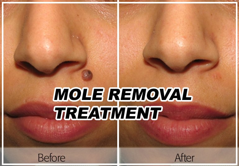
Are you concerned about a mole on your skin? Understanding the symptoms and available treatment options is crucial for maintaining healthy skin. In this article, we will explore the signs and symptoms that may require mole treatment. Additionally, we will discuss effective treatment options that can help address mole-related concerns and ensure your skin’s well-being. Every one of us has moles some hardly visible, some which are our identity features & some bothersome. While most times there is no need to actively remove or treat moles, it is important to differentiate them from viral infections, seborrheic dermatoses &freckles.
Signs and Symptoms
Changes in Size, Shape, or Color:
Moles that grow larger over time or exhibit asymmetry should be evaluated by a dermatologist.
If a mole develops irregular borders or displays uneven coloration, it may require medical attention.
Itching, Bleeding, or Crusting:
Moles that become itchy, bleed spontaneously, or develop a scaly or crusty surface should be assessed by a healthcare professional.
These symptoms could indicate potential skin abnormalities that need treatment.
Pain or Tenderness:
Moles that are painful or tender to the touch should be examined by a dermatologist to rule out any underlying issues.
Rapid Growth:
Moles that experience sudden and rapid growth warrant evaluation by a medical professional.
Monitoring their growth is essential to catch any concerning developments early on.
Effective Mole Treatment Options
Surgical Excision:
This procedure involves removing the mole and a small amount of surrounding healthy tissue.
Surgical excision is commonly used for suspicious or cancerous moles to prevent further complications.
Shave Excision:
Suitable for raised moles, this technique involves using a surgical blade to shave off the mole’s surface.
Shave excision is typically used for non-cancerous moles that cause cosmetic concerns or physical discomfort.
Laser Removal:
Laser technology can be employed to break down the pigmented cells within the mole, effectively removing it.
This method is often preferred for small and non-cancerous moles, leaving minimal scarring.
Cryotherapy:
Cryotherapy involves freezing the mole with liquid nitrogen, causing it to blister and fall off.
This method is commonly used for small, non-cancerous moles and may require multiple treatments.
How do Dermatologists Remove Moles?
Excision (Surgical Removal): For larger or suspicious moles, dermatologists may opt for excision. The area around the mole is usually numbed with a local anesthetic, and the mole is cut out using a surgical scalpel. The wound is then closed with stitches, which may be absorbable or require removal after a certain period of time.
Shave Excision: This method is used for raised moles that are not suspected to be cancerous. The mole is shaved off using a surgical blade, usually at skin level. Stitches are generally not required, and the wound typically heals on its own, forming a small scar.
Laser Removal: Laser technology can be employed to remove certain types of moles. This method is commonly used for smaller, benign moles that are closer to the surface of the skin. The laser targets the pigmented cells within the mole, breaking them down. Several laser sessions may be necessary for complete removal.
Cryotherapy: Cryotherapy involves freezing the mole using liquid nitrogen. The extreme cold destroys the mole’s cells. This method is suitable for smaller moles or flat spots on the skin. Multiple sessions may be required to achieve complete removal.
Electrocautery: In this procedure, an electric current is used to burn and remove the mole. Electrocautery is typically reserved for smaller, benign moles and is often performed with local anesthesia.
Punch Biopsy: A punch biopsy tool is used to remove the entire mole, including deeper layers of skin. This method may be employed if a mole appears suspicious or for diagnostic purposes.
Features of Cancer
When it comes to mole removal, it’s important to understand the features of cancerous moles, also known as melanoma. While I can provide general information, it’s essential to consult with a healthcare professional for an accurate diagnosis and appropriate medical advice. Here are some features or characteristics of cancerous moles:
Asymmetry: Melanoma moles are typically asymmetric, meaning one half of the mole does not match the other half. In contrast, non-cancerous moles are usually symmetrical.
Irregular Borders: Cancerous moles often have irregular or poorly defined borders. They may appear blurry, jagged, or notched, unlike benign moles that tend to have smooth and even borders.
Color Variation: Melanoma moles can display various colors or shades within the same mole. They may include different shades of brown, black, blue, red, or white. Non-cancerous moles often have a uniform color.
Diameter: While melanomas can be small, they generally have a larger diameter compared to benign moles. Although size alone is not a definitive indicator, moles larger than 6 millimeters (about the size of a pencil eraser) are more concerning.
Evolution: Changes over time are significant indicators of melanoma. Cancerous moles may evolve in terms of size, shape, color, or other features. Any new mole or a pre-existing mole that exhibits changes should be evaluated by a dermatologist.
Itching, Pain, or Bleeding: Melanoma moles may be accompanied by symptoms like itching, tenderness, pain, or bleeding. These symptoms should be taken seriously and assessed by a medical professional.
Conclusion
Mole treatment depends on the type of mole and the specific concerns associated with it.Mole removal is a medical procedure aimed at removing moles from the skin. Moles, also known as nevi, are common growths on the skin that can vary in size, shape, and color. While most moles are harmless, some individuals may choose to have them removed for various reasons, including cosmetic concerns, suspicion of malignancy, or discomfort caused by their location or size.Monitoring your moles for any changes and being aware of the symptoms that may indicate the need for treatment is essential for maintaining healthy skin. If you notice any suspicious symptoms or have concerns about your moles, it is crucial to consult a dermatologist. They can accurately diagnose any issues, recommend appropriate treatment options, and ensure your skin’s well-being. Remember, early detection and intervention play a vital role in effective mole treatment and preventing potential complications.